Objective: To sketch the shape of the particle
and state the overall shape and size of a certain particle.
Introduction:
Microscope method is used to analyze the shape and size of a
certain particles. In this experiment, we were given sands with different
sizes, mixed-size sands and lactose to be examined.
Apparatus: microscope
Material: mixed-size
sands, lactose, sand (355 µm), sand (500 µm), sand (850
µm), and slide.
Procedures:
1)
Different types of samples were analyzed by
using microscope, by observing the size and shape of given particle.
2)
The
particle shape is sketched and the overall particle shape of that material is
stated.
Observation:

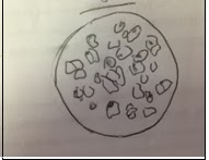
Sand with various sizes
sand (500 µm)

sand (355 µm)

Sand (850 µm)

lactose
Discussion:
In this experiment, the different particle sizes of sand, mixed-size sands and lactose are the chosen materials that are observed. They are observed using light microscope in their place of most minimum orientation, so that determination of shape was made easier and more convenient. However, the specimens prepared should be adequately dispersed on the slide to prevent avoid agglomeration that will affect the observation. All the particles of sand observed are in irregular shape meanwhile lactose is more regular in shape.
Questions:
1)
Briefly
describe the various statistical methods that can be used to measure the
diameter of a certain particle.
There are four statistical methods
that can be used to measure the diameter of a certain particle: Firstly,
Projected area diameter which is based on the equivalent area to that of the
projected image of a solid particle. Secondly, projected perimeter diameter
which is based on the circle having the same perimeter as the particle. These
two methods can be carried using light microscope. Thirdly, Feret’s diameter
that is the mean distance between two parallel tangents to the projected
particle perimeter. Forthly, Martin’s diameter that is the mean chord length of
the projected particle perimeter, which can be considered as the boundary
separating equal particle areas. These two methods are used for three-dimensional
image by using electron microscope. However, the values are dependent on both
the orientation and the shape of particles.
2)
State
the best statistical method for each sample that you used.
For all materials that we used, the
best statistical method is Feret’s and Martin’s diameter. It is because both of
these are the statistical diameter which is averaged over many different
orientations to produce a mean value for each particle diameter.
Conclusion:
Generally lactose particles appear to be smaller than the
sand particles that have been used in this experiment. Different types of
particles have different shapes. Particle size should be taken into
consideration during the production of drug with appropriate dosage forms
because particle size will influence pharmacodynamics and pharmacokinetics of
the drugs in body.
References:
1)
Aulton,
M.E. (2002). Pharmaceutics: The science of dosage form design. Edinburgh:
Churchill Livingstone.
2)
Physicochemical
Principals of Pharmacy (2nd
Edition) AT Florence and
D.Attwood The MacMillan Press Ltd
No comments:
Post a Comment